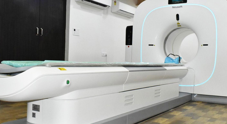
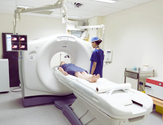
Advanced nuclear imaging methods like positron emission tomography (PET) scans are used to check for cancer and its spread. PET scanners can track the absorption of a radioactive sugar by the body’s cells. Cancer cells use more sugar (glucose) than normal cells due to their rapid growth. Before the test, the patient will either drink or receive an injection of the sweet tracer. The patient lies on a table during the nuclear medicine scan and then enters a sizable scanner with the shape of a tunnel. The length of the outpatient process varies based on the area of the body being examined and is painless.
What is a PET/CT scan?
This cutting-edge method of nuclear imaging combines a computed tomography (CT) scan with a PET scan in one device. Similar to conventional X-rays is a CT scan. But it captures images from various perspectives in tiny segments. These tiny slices are assembled by a computer to provide a 3D image of the X-rayed region. In a single session, the use of CT and PET imaging provides information on the structure and function of cells and tissues throughout the body (from the CT scan). A PET/CT scan gives medical professionals the ability to study diseases and abnormalities at the cellular level, as opposed to an X-ray or magnetic resonance imaging (MRI) scan.
PET scan vs. CT scan
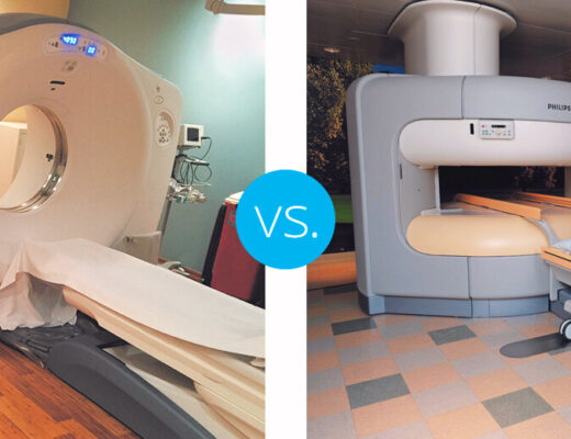
Whereas CT scans take pictures from a variety of angles to show images of the patient’s body organs, tissues and bones, the PET scan shows how the patient’s cells react to a radiotracer, which may indicate cancerous areas. The combined PET/CT scan joins these two technologies together.
What is PET/MRI?
A PET/MRI scan is used to obtain anatomic and quantitative information from an MRI at the same time as physiologic information from PET imaging. The advantage of a PET/MRI is that it shows soft tissue more clearly. Soft tissue cancers occur most frequently in connective tissue and are often found in the legs and pelvis. They may also develop in the arms and upper body.
What does a PET/CT scan show?
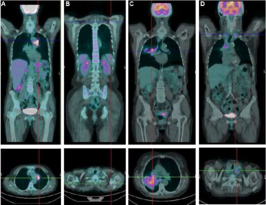
Damaged or malignant cells that are being taken up by the radiotracer mixture can be “seen” by the PET/CT scanner. In some cancers, the pace at which the tumour utilises the sweet ingredient may aid in determining the tumour grade. By identifying which areas of the body have aberrant activity, this aids in staging your tumour.
A PET/CT scan offers a more thorough image of malignant tissues than each test by itself because it combines information about the body’s structure and metabolic activity. A single scan produces both the PET and CT pictures, which allows for great accuracy. The oncology team can create the best cancer treatment strategy with the aid of a PET/CT scan. A PET/CT scan may also:
- Information about the effectiveness of a treatment should be provided.
- help with future radiation therapy planning
- Find the appropriate location on the body to perform a biopsy, if necessary.
- During follow-up care, once treatment is finished, look for any new cancer development.
Compared to other imaging procedures, a PET/CT scan has the potential to be more sensitive and might detect cancer earlier. Even while not all tumours are radiotracer-receptive, PET/CT is quite accurate in differentiating between benign and malignant tumours when it detects them, especially in specific malignancies like lung and musculoskeletal tumours.
How to prepare for a PET/CT scan
Look for the ideal facility for you: The radiology or nuclear medicine department of a hospital normally performs PET/CT scans, but you might also be able to schedule an appointment at an outpatient imaging centre.
- Dress appropriately: Wearing loose clothing could be more practical because you might be asked to undress for the treatment. The establishment could provide you a shawl or gown to wear. Additionally, it’s possible that you’ll be asked to take off any jewellery, piercings, or metal accessories like dentures or hearing aids.
- Bringing your medical records Your personal and medical history, including previous scans and operations, as well as a list of the current drugs you’re taking, may be requested by the technician.
- Review the guidelines for eating and drinking: You might be instructed to go without meals for around six hours before to the PET/CT scan and to only consume water. Before the operation, consult your doctor because this may vary from instance to case. Although there isn’t a set diet for PET scans, eating can affect the radioactive tracer’s distribution, resulting in less than ideal results. Avoid coffee and sugary beverages. The metabolism of sugar (glucose) can be impacted by caffeine in coffee.
- Enquire about exercise Exercise should be avoided 24 to 48 hours before to the PET/CT scan, per your doctor’s instructions.
- Give yourself ample time since a little quantity of a radioactive sugar material, which takes 30 to 90 minutes to travel throughout the body, will be administered to you to aid the scan in producing its photos. 30 minutes or so are needed for the scanning process. The length of time depends on the bodily part that has to be scanned. Spend one to three hours at the imaging facility.
- Lean on a friend or member of your family: It could be beneficial to bring a loved one with you if you suffer from anxiety. Find out from the institution if there are any limitations on bringing assistance.
During the scan
Be ready for the position: You could be requested to lie down on a table that slides into a big scanning device that has a hole in the centre shaped like a doughnut. The technician might urge you to hold your breath sometimes throughout the scan in order to acquire quality pictures.
A shot is anticipated: Through an intravenous (IV) line, a little quantity of a radioactive sugar material is administered into your bloodstream. Cancer is drawn to and absorbs the energy of this material, making it easier for the scanner to find it and create photos of the interior of your body.
Do not be averse to speaking out: Tell the technician if you’re feeling claustrophobic or nervous before the PET/CT scan because these feelings are typical.
After the scan
- Resume activities: Typically, you may resume normal activities right after the scan.
- Drink water: It’s a good idea to drink plenty of water afterward to flush out the dye or radioactive sugar.
- What are possible side effects and risks of a PET/CT scan?
- While the benefits of a PET/CT generally outweigh the risks, there are some risks.
What do the results mean?
A radiologist who specializes in reading PET/CT scans will interpret the images and report back to the doctor who ordered the procedure. The radiologist’s report will include:
- Whether your scans show signs of cancer (and if there is cancer, what stage it is)
- Whether the cancer has spread and where
How long it takes for your doctor (the ordering physician) to get your results depends on a number of factors:
- Did your doctor request immediate results? Is there an urgency?
- How complex is your particular test?
- Has your doctor given the radiologist all the information needed to interpret the images?
- Is this your first PET? If you had prior scans, the reader may need to see them for comparison. Does the reader have your previous scans or need to get them?
- How is the report from the reader being sent: phone, email, fax or regular mail?
- Don’t be afraid to ask the facility when your doctor is likely to receive the report.
Conclusion
The radiotracer can cause an allergic response in you. A modest allergic response is typical. Beforehand, inform your care staff of any allergies. If the radiotracer is injected into you, you can feel a little discomfort. It ought should disappear shortly. As the contrast is administered, you can feel warm or flushed.
Also See:


