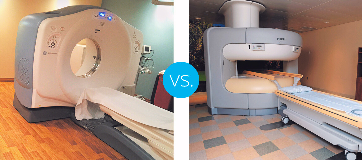
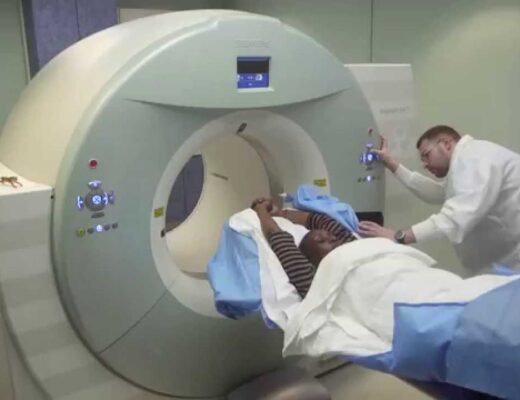
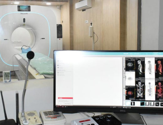
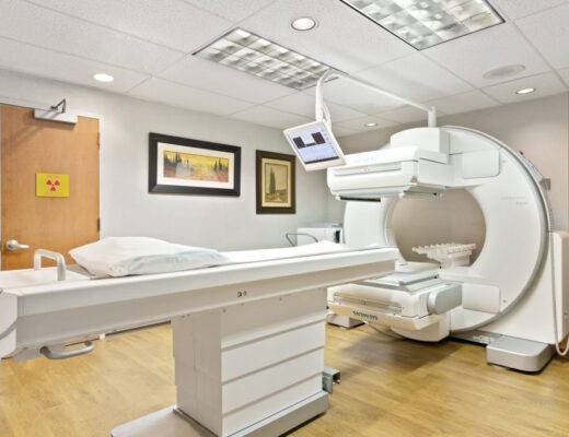
In the realm of modern medicine, understanding the complexities of the human body often requires a peek under the hood with advanced imaging techniques like PET-CT scan and MRIs. Both are highly effective tools, each with strengths in distinct areas. Let’s delve into their key differences to help navigate your imaging decisions.
PET-CT Scans: Unveiling Cellular Activity
PET (Positron Emission Tomography): This technique uses a small amount of radioactive tracer injected into your body. The tracer accumulates in areas with high metabolic activity, like growing tumors or regions of inflammation. A special camera detects the emitted radiation, creating images that reveal cellular function.
CT (Computed Tomography): A PET scan is often combined with a CT scan for anatomical detail. The CT scan uses X-rays to create detailed cross-sectional images of your organs, bones and tissues. By combining the functional information from the PET scan with the anatomical detail of the CT scan, doctors get a clearer picture of what’s happening inside your body.
When is a PET-CT Scan Used?
- Cancer diagnosis and staging: PET-CT scans excel at detecting cancerous tumors due to their increased metabolic activity. They also help determine the stage of cancer, revealing if it has spread to other parts of the body.
- Heart disease evaluation: PET-CT scans can assess blood flow to the heart muscle, identifying areas with potential blockages.
- Neurological disorders: PET-CT scans can help diagnose conditions like Alzheimer’s disease and Parkinson’s disease by visualizing changes in brain activity.
MRI Scans: Unveiling Structure and Anatomy
MRI (Magnetic Resonance Imaging): This method utilizes powerful magnetic fields and radio waves to generate precise images of organs, soft tissues and bones. Unlike PET scans, MRIs do not expose patients to radiation.
When is an MRI Scan Used?
- Soft tissue injuries: MRIs excel at visualizing muscles, ligaments and tendons, making them ideal for diagnosing sports injuries or tears.
- Musculoskeletal issues: MRIs are valuable for examining bones, joints and the spine, aiding in the diagnosis of conditions like arthritis or spinal disc herniations.
- Brain imaging: MRIs offer precise images of the brain, aiding in diagnosing tumors, strokes and other neurological disorders.
The Final Verdict: A Collaborative Approach
Choosing between a PET-CT scan and an MRI depends on your specific needs. Here’s a simplified breakdown:
- For assessing cellular activity and function: PET-CT scan.
- For detailed anatomical visualization of soft tissues, bones and organs: MRI scan.
Conclusion
According to rnmcenter PET-CT scans are best for evaluating cellular activity in conditions like cancer and heart disease, while MRI scans provide detailed images of soft tissues and organs, essential for diagnosing musculoskeletal and neurological issues. Combining both techniques often provides a comprehensive view for precise diagnosis and effective treatment planning.


