PET-CT in Neurology: Detecting Dementia and Brain Disorders Early
🧠 Introduction: Looking Inside the Living Brain
When it comes to understanding the brain, few imaging technologies have been as revolutionary as PET-CT scanning.
At RNM Centre for Nuclear Medicine & Molecular Imaging, Rohtak, PET-CT plays a crucial role in diagnosing neurological disorders — from Alzheimer’s disease and epilepsy to movement disorders and brain tumors — often before structural changes appear on MRI or CT scans.
Unlike traditional imaging, PET-CT doesn’t just show what the brain looks like — it reveals how it functions. This ability to visualize metabolic and molecular activity makes it indispensable in modern neurology.
⚛️ What Makes PET-CT Unique in Brain Imaging
PET-CT combines two powerful modalities:
- Positron Emission Tomography (PET) detects how brain cells consume energy (usually glucose).
- Computed Tomography (CT) provides a detailed map of brain structure.
When combined, they reveal areas of the brain that are overactive, underactive, or damaged, helping clinicians detect disease at its earliest stage.
💡 How PET-CT Works for Brain Evaluation
- Tracer Injection:
A safe, low-dose radioactive glucose compound called Fluorodeoxyglucose (FDG) is injected into the bloodstream. - Tracer Uptake:
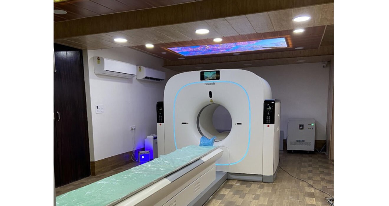
Brain cells absorb FDG in proportion to their activity level. - Imaging Process:
The PET-CT scanner captures signals from the tracer, generating a 3D image of glucose metabolism across brain regions. - Analysis:
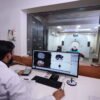
Nuclear medicine experts at RNM Rohtak interpret these patterns to detect abnormalities in brain function — even before symptoms become advanced.
🧬 Key Neurological Conditions Diagnosed with PET-CT
1. Alzheimer’s Disease and Dementia
- Detects reduced glucose metabolism in the temporal and parietal lobes.
- Helps differentiate Alzheimer’s from other dementias such as frontotemporal or vascular dementia.
- Aids in early diagnosis — when cognitive symptoms are mild or unclear.
- Assists doctors in tracking disease progression and therapy response.
2. Epilepsy Evaluation
- Identifies seizure foci by locating areas with decreased metabolism between seizures (interictal phase).
- Crucial for patients being considered for epilepsy surgery, ensuring precise localization of seizure origin.
3. Parkinson’s Disease and Movement Disorders
- PET scans with specific tracers like F-DOPA or DAT (dopamine transporter) ligands help distinguish Parkinson’s disease from other movement disorders such as multiple system atrophy or essential tremor.
4. Brain Tumors and Post-Treatment Assessment
- Differentiates between tumor recurrence and radiation necrosis after therapy.
- Evaluates tumor metabolism to guide biopsy and treatment planning.
5. Psychiatric and Metabolic Brain Disorders
- Provides insight into depression, schizophrenia, and autism spectrum disorders by visualizing altered neurotransmitter activity.
- Assists in research and personalized neuropsychiatric therapy.
🌟 Why Early Detection Matters
| Stage | Traditional MRI Findings | PET-CT Findings |
| Early / Pre-symptomatic | Often normal | Detects reduced metabolism |
| Mild Cognitive Impairment | Subtle or absent changes | Distinct hypometabolic patterns |
| Advanced Disease | Structural loss | Confirms and quantifies severity |
Early PET-CT evaluation allows neurologists to start treatment early, manage progression, and provide families with timely counseling and care options.
🛡️ Safety and Comfort
PET-CT brain scans are non-invasive, safe, and painless.
At RNM Rohtak:
- The tracer dose is minimal and fully regulated by AERB guidelines.
- Radiation exposure is comparable to standard diagnostic imaging.
- The entire procedure, including preparation, takes about 90 minutes.
- Patients can return home the same day.
🚀 The Future of Brain Imaging
Modern advancements in molecular PET tracers — such as Amyloid and Tau imaging agents — can now visualize the actual protein deposits that cause Alzheimer’s disease.
These technologies, when combined with AI-based analytics, are bringing us closer to personalized brain health assessments.
At RNM Rohtak, we are proud to offer next-generation PET-CT imaging that supports neurologists in delivering early, precise, and effective care for patients with brain disorders.
🏥 About RNM Rohtak
RNM Centre for Nuclear Medicine & Molecular Imaging, Rohtak, is one of North India’s most advanced centers for PET-CT, SPECT-CT, and radionuclide therapies.
Our expert team uses molecular imaging to diagnose neurological, oncological, and cardiac diseases with the highest precision and compassion.